18+ Eye Structure Diagram Blank Gif. It is situated on an orbit of skull and is supplied by optic nerve. Is a health blogger focusing on health beauty lifestyle and fitness topics.
Blank Eye Diagram - Cliparts.co from cliparts.co Download blank eye diagram and use any clip art,coloring,png graphics in your website, document or presentation. Learn vocabulary terms and more with flashcards games and other study tools. This article is about blank eye diagram.
Image of 14 best human eye drawing images art drawings drawings.
Please click on the image(s) to view larger version. Feel free to browse our website for best viewed on 1280 x 768 px resolution in any modern browser. Lucidchart helps users sketch and share professional flowchart diagrams, providing designs for anything from brainstorming to project management. Blank diagram of the eye blank eye diagram studioy see more.
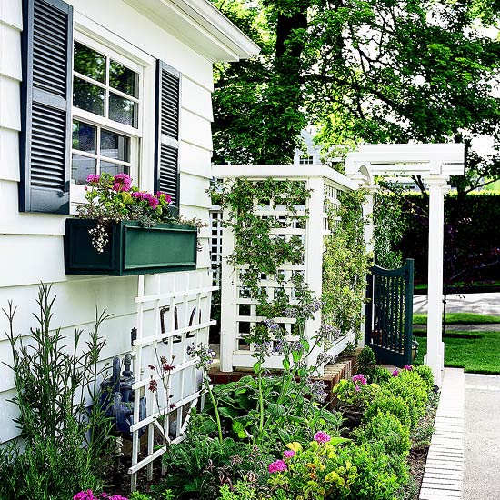
36+ Trellis Design Pics . Handmade dolls and clothing (no custom orders) all images and doll designs remain the intellectual property of trellis. Visit again soon to see what amazing website they decide to build. Open Shelter from 4.bp.blogspot.com But as time goes by the trellis also functions as a decoration of houses, buildings and so on. This structure is designed to give climbing plants room to grow. If you grow indeterminate tomatoes, these tomato trellis designs and ideas will make you head on out to the hardware store and get to building! Trellis & pergola design ideas for landscape design. Alibaba.com offers 6,832 design trellis products. If you do not have a lot of square footage for gardening, try using trellises or posts in your garden. Pyramid trellises, likewise at times alluded to as monoliths, make amazing central focuses in a garden or scene plan. Trellis design v...
46+ Trellis Stardew Valley Background . Leave me a comment with your feedback, would love to hear what you think and tips on how i can improve!follow me on twitter for updates and more. Trellis are wooden frameworks that help certain plants (for example beans) with growing. How to get Green Beans - Stardew Valley - YouTube from i.ytimg.com Leave me a comment with your feedback, would love to hear what you think and tips on how i can improve!follow me on twitter for updates and more. In stardew valley their main effect is, that plants who need trellis block your movement. Rated 4.84248 from 38,351 votes. Stardew valley scarecrow walk through trellis scarecrow range stardew quality sprinkler stardew stardew valley best farm stardew valley crops stardew valley trees stardew valley layout. Trellis are wooden frameworks that help certain plants (for example beans) with growing. A couple of...
Get Anatomy Of Ct Scan Brain Background . Brain bones of cranium sinuses of the face. Brain ct scans may be done with or without contrast. contrast refers to a substance taken by mouth or injected into an intravenous (iv) line that causes the particular organ or tissue under study to be seen more clearly. CT Scan Illustration Depicting Normal Brain Anatomy and ... from www.patientedlibrary.com The video shows the basic ct anatomy of the brain.for each slice we have highlighted. The ct head scan is one of the most common imaging studies that you can be faced with and the most frequently requested by a&e. Anatomy of the head on a cranial ct scan : The ct head scan is one of the most common imaging studies that you can be faced with and the most frequently requested by a&e. Contrast examinations may require you to fast for a certain period of time before the. Learn about brain an...


Comments
Post a Comment